
Ultra-fast Cooling Technology
Please see the background introduction in the Cell and Tissue Cryopreservation page. Different from traditional cryopreservation technologies, vitrification without any type of cryoprotectant, including both cell permeating (e.g. DMSO and glycerol) and non-permeating types (e.g. saccharides, polymers, or antifreeze protein analogues) would be ideal for cryopreservation of thin tissues or tissue-like structures.
This “in-situ” cryopreservation approach significantly simplifies the cryopreservation protocol, eliminates toxicity and other negative impacts incurred by using cryoprotectants (e.g. osmotic damage, unwanted differentiation of stem cells and contamination of animal products). Theoretically, this outcome can be achieved by employing an ultra-fast cooling rate (106 K/min required for cell suspensions and possibly 105 K/min for dense tissues which possess much less extracellular water). Currently, the methods generally practiced by traditional vitrification procedures involve directly plunging samples into cryogenic fluid (e.g. liquid nitrogen or helium). Those methods cannot achieve the above cited cooling rates required for direct vitrification, in large part because the procedures cited result in strong vaporization around the sample surface, and this action will form a “vapor coat” which acts as a heat-insulation layer. As a result, the heat transfer coefficient between the sample surface and liquid nitrogen is quite limited due to poor thermal conduction across the “vapor coat”. This limitation prevents a further increase of the cooling rates.
Recent development of ultra-fast cooling technologies using nitrogen slush (mixing liquid and solid nitrogen) and swinging thin quartz straws, metal meshes for high pressure freezing, micro chambers moving at high speed for injection into liquid nitrogen, etc., have significantly reduced the cryoprotectant concentrations and achieved promising progress toward to the ultimate goal of direct vitrification. However, application of these technologies is limited to miniature sample sizes, typically on the order of several microliter or even several hundred nanoliters). Some of the methods also require high velocity movement of miniature samples (e.g. swinging and ejection) or high pressures, which are obviously impractical for tissues.
CryoCrate’s InstaVitria® technology, see Technology in more details, is based on an advanced thermal fluidic design to prevent the formation of vapor coat on sample surface. More importantly, the design achieves uniform distribution of the ultra-fast cooling rate on a relatively large area that is sufficient to cover the surface of artificial tissues (e.g. stem cell derived artificial tissues) and some biological tissues (e.g. corneas, relatively small human skin grafts and small plant tissues). This system under R&D potentially achieves in-situ cryopreservation for those tissues that are highly valuable for both academic research and biomedical applications and benefit highly valuable markets, such as tissue engineering and drug discovery using artificial tissues.
Another application of using InstaVitria® technology is to prevent cell ultra-structure damage during cryopreservation. For example, traditional cryopreservation methods cause irreversible damage to intracellular lipid vesicles of intact pig embryos. Facing the fast advance of the genetic editing and engineering technologies and xenotransplantation applications, efficient cryopreservation of pig embryos will be soon be in high demand for numerous associated industries. Traditional cryopreservation technologies result in poor survival rate for pig embryos unless the intracellular lipid is removed, but the intracellular lipid is critically important for further embryonic development. As such, there are currently no efficient strategies for long-term storage of pig embryos, which is in sharp contrast to that for bovine embryos which result in transfer of over 200,000 frozen embryos worldwide.
A variation of CryoCrate InstaVitria® system shows promising results in preventing deformation of intracellular lipid vesicles of pig embryos during cooling, and has the potential in opening numerous new market opportunities for banking of pig embryos for numerous agricultural and biomedical applications.
Cooling Artificial Tissues with One Layer of Mesenchymal Stem Cells Without Any Cryoprotectant and With or Without Using InstaVitria®
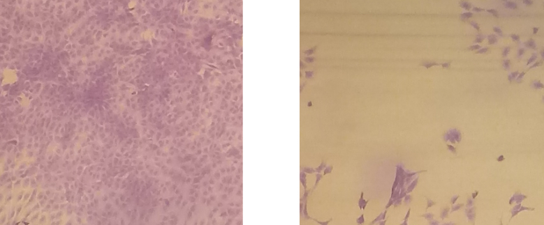
A comparison between the morphology of the layer of mesenchymal stem cells on artificial tissues treated by InstaVitria® system (LEFT) using the hermetic tissue holder designed for the system and frozen by plunging into a liquid nitrogen pool (RIGHT) and using the same tissue holder. No cryoprotectant was used. The tissue holder has a special ultra-thin steel cover (20 μm in thickness) to prevent direct contact between the cell layers with non-evaporating liquid nitrogen jets from the InstaVitria® system or with the boiling liquid nitrogen in the cooling pool. The cell layer portion processed by the InstaVitria® ultra-fast cooling approach maintained the colony integrity and high viability, while that frozen by liquid nitrogen pool boiling lost almost all cells.
Cooling Pig Skins With or Without Using InstaVitria®
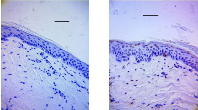
Comparison between the post-thaw viabilities of porcine skin tissues cryopreserved using InstaVitria® technology (LEFT) and slow freezing technology (RIGHT) based on TUNEL staining with dark brown illustrating apoptosis (bar = 100 µm) for various cell types.
Vitrification of Pig Intact Embryos With or Without Using InstaVitria®
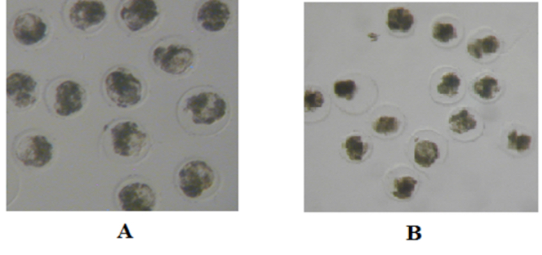
Significant improvement of porcine embryo (blastocytes) post-thaw morphology by preventing the intracellular deformation with InstaVitria® (A), compared to traditional vitrification protocol (B).